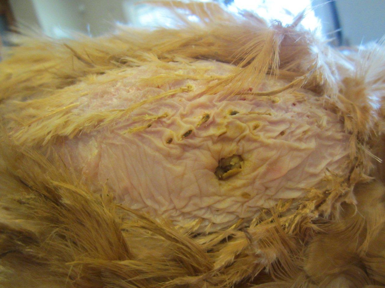
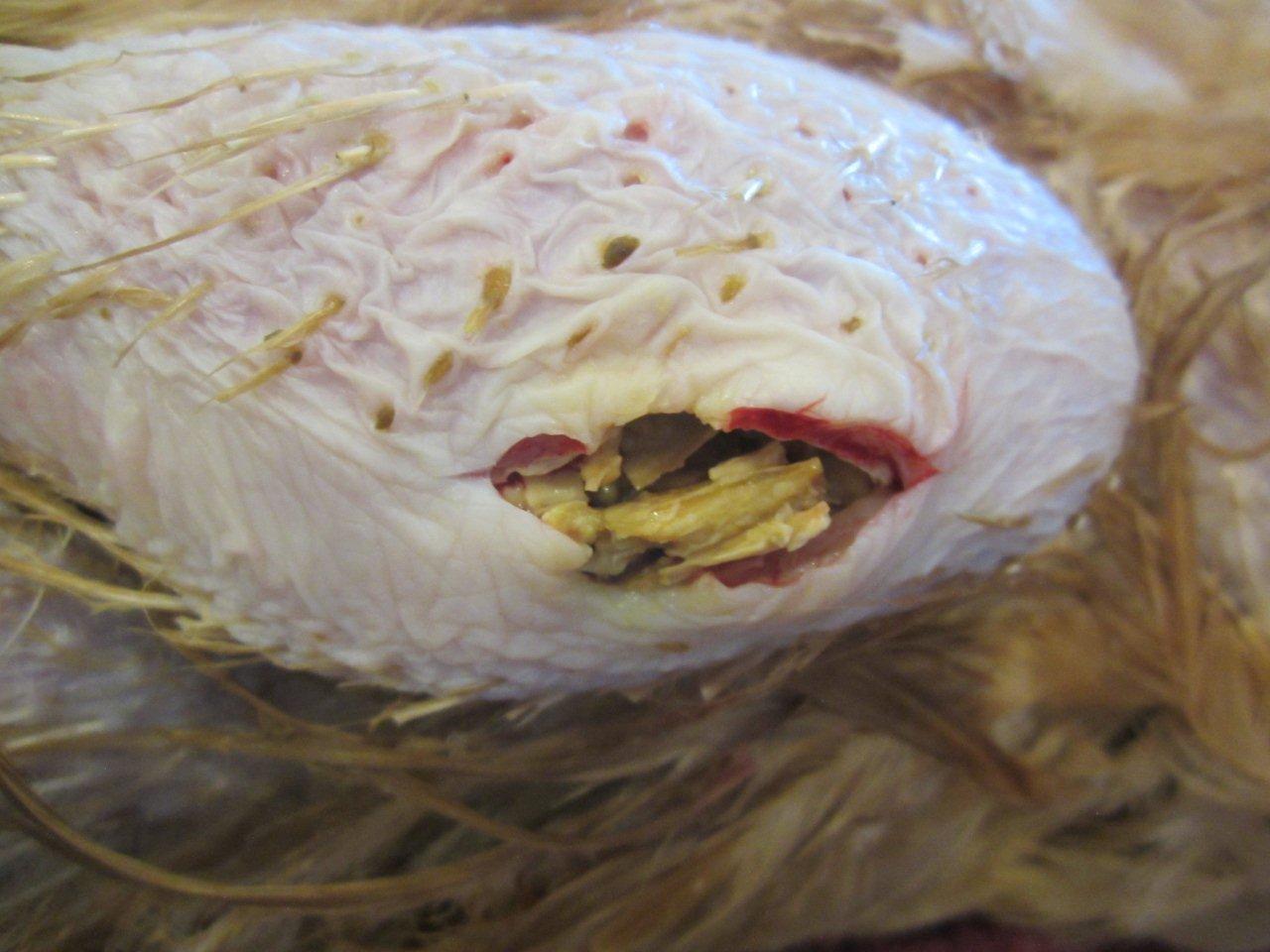
I’m assuming this is for @windychicks for reference to her chicken with an unidentified tumor/abscess? Looks like her bird has more of a tumor looking lump than an abscess like the above pictured.
I’m assuming this is for @windychicks for reference to her chicken with an unidentified tumor/abscess? Looks like her bird has more of a tumor looking lump than an abscess like the above pictured.
Chickens can get all sorts of tumours, some of which are caused by Marek’s disease (herpes virus) and others by mainly viral causes, particularly retroviruses. The chicken has the notoriety of being the first animal discovered with cancer caused by a retrovirus, this was in the 1930s. Tumours can be found almost anywhere in the chicken as lymphoid tissue is spread throughout the bird (unlike mammals which have lymph nodes). The main viruses, apart from Marek’s, are the avian leukosis/sarcoma viruses, plus squamous cell carcinoma (SCC).
How common?
Tumours caused by the Marek’s virus are the most common (the acute form is where birds may die suddenly with no symptoms and tumours may be found in the liver, gonads, spleen, kidneys, lungs, proventriculus, heart, muscle and skin). Retroviral tumours are less common (see below). Tumours in the skin (SCC) are occasionally seen. Retrovirus survival outside the body is only hours, so the disease is not very contagious to other members of a flock.
Symptoms
Due to the general spread of lymphoid tissue (where cancer cells grow the fastest) in a chicken, lethargy, lack of appetite and dullness are the most common symptoms with possibly diarrhoea, pale wattles and an enlarged liver. The lymphoid leukosis virus slowly transforms cells into neoplastic ones and the acutely transforming retroviruses do the damage faster, all of them using and damaging genes and causing tumours which can include fibrosarcoma, chondroma, endothelioma, haemangioma, nephroblastoma and hepatocarcinoma – affecting potentially almost all of the organs in the chicken’s body. The incubation period of lymphoid leukosis from infection to the developed disease and death is about four months. Losses occur from 5-9 months of age in egg-laying and breeding stock. Other leukosis viruses affect adults sporadically as the virus is ubiquitous in poultry worldwide and is passed down through the egg as well as transmitted by direct or indirect contact. Lymphoid leukosis (most common): enlarged liver, spleen, bursa, kidneys, ovary.
Erythroid leukosis (rare): moderately enlarged liver and spleen, leukaemia, liquid cherry red bone marrow.
Osteopetrosis (rare): thickening of long bones in legs and wings.
SCC can occur anywhere on the skin but is most commonly seen around the face and eyes with the subsequent swelling often being be mistaken for respiratory infection or an abscess. Sometimes, feather follicles can be affected.
Treatment
Unfortunately, no treatment or vaccines are available for leukosis. Chemotherapy tends to be too toxic to birds to be useful. Control is based on high standards of hygiene and selection for resistance to the viruses by culling affected birds. The diagnosis via your vet and laboratory is by virological testing, post mortem lesions and the presence of tumours. The Marek’s vaccine mostly controls the disease.
Prevention
Some breeds are resistant to tumour development, such as the Fayoumi. Good husbandry with minimal stress is always useful to maintain healthy chickens. Removing their progeny from affected adults will help reduce the viral shedding as the virus is passed down through the egg.
Vaccination
None has been developed as these retroviruses are not sufficiently common to cause industrial poultry problems. However, we do as backyard poultry keepers benefit from many vaccines that the poultry industry has developed to ensure healthy food production for us. The Marek’s vaccine is fairly well known, used in the industry and by keepers of Silkies and Sebrights.
I found the following here: http://avianexoticsvet.com/case-of-the-month/#collapse_3_queenie-the-peahen
Queenie, a 4-year-old female peahen, came to the Veterinary Center for Birds & Exotics because of her swollen eye. A peahen is a female peacock; collectively, males and females are called peafowl. Upon first glance, anyone could tell there was something wrong! There was a very large swelling, about the size of a super-sized gumball, in the inner corner of her left eye (see photo #1). The swelling was so large that it was difficult to tell if the eyeball was even there, but after close examination, the doctors determined that the eye was just fine. The sinus that sits just in front of the eye was actually where the problem was.
To determine the cause of the problem, the doctors inserted a needle into the swelling to collect cells to determine if the swelling was an infection or cancer. The sample they took was placed on a microscope slide and submitted to a specialized veterinarian called a pathologist for identification of the cells. The swelling turned out to be an abscess (a pocket of pus) in the bird’s infra-orbital sinus (the sinus in front of the eye).
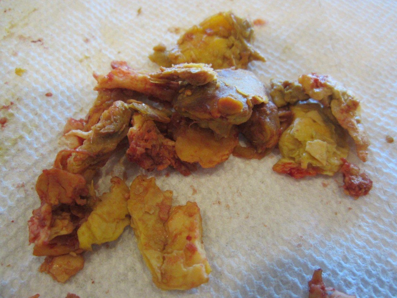
The tricky thing about birds’, herbivores’ (rabbits, guinea pigs, rats), and reptiles’ pus is that it is thick, like cottage cheese or toothpaste, because these animals lack the ability to break it down into a liquid, like our pus. This is the reason we have to perform surgery to remove the pus from these animals and not just cut the area open and let it drain out.
The doctors performed surgery to remove the giant ball of pus from the corner of Queenie’s eye. At first, she was sedated and given pain medication to ease her into anesthesia. Then a breathing tube was placed in her trachea to help us ensure she was able to breathe under anesthesia. The veterinary technicians placed a heart monitor on her to follow her heart rate. Then the area where the surgical incision was to be made was scrubbed with a sterile surgical preparation.
The eye is an area of the body that contains many nerves and blood vessels and has the potential to bleed significantly in surgery. Therefore, the doctors used a specialized surgical tool called an electro-cautery to make the incision, allowing the blood vessels to be cauterized instantly to prevent blood loss. Once the cut through the skin was made, the vets immediately saw thick yellow pus underneath. Very carefully, they loosened the ball of pus from the sinus pocket and removed as it one big solid chunk. Then they flushed the sinus with sterile saline to remove any excess infectious debris. They left the skin incision open so that the sinus could be flushed with sterile saline daily over the following days to try to prevent recurrence of infection. Since the eyes and nose connect via the sinuses, her nostril and the slit in the roof of the bird’s mouth, called the choana, had debris in them, as well; so the doctors cleaned these areas out, too, during surgery. They had to be sure to remove all the pus from the mouth and nose to try to prevent the infection from lingering.
Immediately after surgery, Queenie felt so much better! She could now breathe through her nose again and see out her left eye (see photo #2). Queenie stayed in hospital for the next few days so that the doctors could flush the surgical site. By the time she went home, the incision had already healed all on its own without the use of stitches. She was placed on antibiotics and pain medications to help with the remaining swelling. Today Queenie is doing great! The eye infection hasn’t returned, and she is back with her flock, living her happy peahen life, once again.
Source: http://avianexoticsvet.com/case-of-the-month/#collapse_3_queenie-the-peahen